
Neuronal imaging
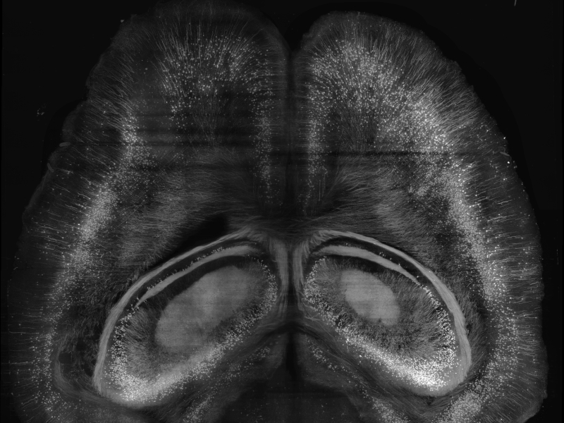
Cross-section of a whole Thy1-GFPM mouse brain imaged with a Light-Sheet Fluorescence Microscope
Description
The neuronal imaging group at INO performs research in high-resolution microscopy techniques for the investigation of biological tissues, with a special interest in neurological samples. In particular the group has gained considerable expertise in designing and building Light Sheet Fluorescence Microscopes and Two-Photon Fluorescence Microscopes. Using these advanced microscopy techniques, it is now possible to explore biological tissues at an unprecedented scale. Whole organs, such as a whole mouse brain, can be imaged with sub-micron resolution thus enabling high-resolution reconstruction of the biological structures on a macroscopic scale. Research within this group also covers two related areas that are essential for the success of these microscopy techniques: biochemical protocols for sample staining and optical clearing, big data management and image processing. The SWITCH and CLARITY/TDE techniques were developed at INO as clearing procedures to render samples transparent to light, hence enabling light to penetrate deep into the samples. On the other hand, given the high resolution and correspondingly huge datasets produced by the microscopes (several terabytes per day), the group has developed knowledge and methods for big data management including volumetric image stitching, compression and automatic feature extraction using artificial neural networks. All this expertise lies at the base of INO�s participation in several important scientific endeavours such as the Human Brain Project, co-funded by the European Commission. In perspective, the know-how gained so far will be used to tackle the present and future challenges of biomedical investigation: scaling up to whole human brain imaging.
INO Staff
Baghdad MohamedCostantini IreneGiannoni LucaMazzamuto Giacomo (Contact Person)Montagni ElenaPavone Francesco SaverioRamazzotti JosephineSilvestri Ludovico
